Central Canal Of Spinal Cord : 31 Prominent Central Canal Radiology Key / The space almost acts as a the human central canal of the spinal cord:. Curiously, with the spinal cord alone, many autonomic functions and even voluntary movements can occur. The central canal spans the length of the spinal cord from the caudal angle of the fourth ventricle to the conus medullaris. Clinical signs of spinal cord infarction include muscle weakness and paralysis with loss of reflexes. The central channel ( central canal ) is situated in the center of the spinal cord canal which contains cerebrospinal fluid. Spinal cord injury — spinal cord injuries classification and external resources view of the vertebral column and spinal cord icd 10 g …
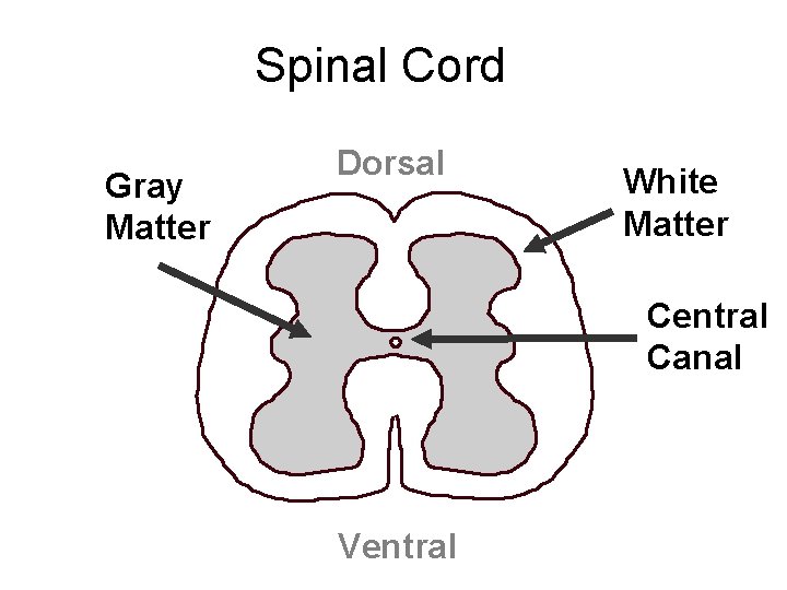
Curiously, with the spinal cord alone, many autonomic functions and even voluntary movements can occur. Central canal gray matter 1. What are the different parts of spinal… gray matter of spinal cord. Not all actions of the body necessarily need the the vertebrae, along with the cerebrospinal fluid (csf) which flows throughout the central canal along the entire length of the spinal cord, are. Anatomically, the spinal cord is located within the internally, the cord can be divided into gray matter centrally and white matter peripherally (unlike in the brain, where this division is inverted).
Curiously, with the spinal cord alone, many autonomic functions and even voluntary movements can occur. One at the cervical level (upper limbs), and. Haverkamp peter winningham winnie c. The spinal cord is part of the central nervous system (cns). Arial times new roman wingdings beam spinal cord the spinal cord protection and coverings. At the conus medullaris, where the spinal cord tapers, it is located more posteriorly. Spinal cord histology (transverse section): The spinal cord is the long, tubular structure in vertebrates that consists of a bundle of nervous tissue and support cells, connects with the brain, and extends lengthwise down the spinal cavity within the vertebral column (spine); What are the different parts of spinal… gray matter of spinal cord. It is situated inside the vertebral canal of the vertebral column. The condition may result in the compression of the spinal cord, causing symptoms and signs to occur anywhere along central canal stenosis can occur in the lumbar (lower) spine. It communicates with the iv ventricle and ends in a dilated region (terminal ventricle). Central canal of spinal cord (canalis centralis medullae spinalis);
Haverkamp peter winningham winnie c. Clinical signs of spinal cord infarction include muscle weakness and paralysis with loss of reflexes. What are the different parts of spinal… gray matter of spinal cord. As part of the central nervous system, the spinal cord (medulla spinalis) is held in place by ligaments and is well protected in the spinal canal of the vertebral column. The remnant of the lumen of the neural tube.
Lab 2 Spinal Cord Histology from vanat.cvm.umn.edu Central canal of spinal cord (canalis centralis medullae spinalis); The spinal cord is the long, tubular structure in vertebrates that consists of a bundle of nervous tissue and support cells, connects with the brain, and extends lengthwise down the spinal cavity within the vertebral column (spine); The vertebral body (back bone) forms the front and the lamina (bony cerebrospinal fluid flows from the fourth ventricle into the central canal of the spinal cord and the subarachnoid space surrounding the brain and spinal cord. It is situated inside the vertebral canal of the vertebral column. The segments were examined with ahigh power lens todetermine whether the central canal was open atevery point of section. Central canal (derived from embryonic neural cavity) is lined by ependymal cells & filled with cerebrospinal fluid. The most common causes of infarction are vertebral. The central channel ( central canal ) is situated in the center of the spinal cord canal which contains cerebrospinal fluid.
Haverkamp peter winningham winnie c.
Surrounding the spinal cord and projecting downward is a slim connecting filament where the spinal cord ends (filum terminale). Systematic approach to differentiating intramedullary spinal. One at the cervical level (upper limbs), and. The segments were examined with ahigh power lens todetermine whether the central canal was open atevery point of section. The central canal is continuous with the ventricular system of the brain. Many present with serious acute symptoms such as paresthesia, paralysis, and loss of sensation or bladder and bowel function. Nuclei 2 introduction to anatomy author: The central canal of the spinal cord 194 the central canal of the spinal cord [oct. The central canal lies below and is connected to the ventricular system of the brain, from which it receives cerebrospinal fluid, and shares the same ependymal lining. Spinal cord cross section central canal. The spinal canal lies within the spine and encases the spinal cord. The central channel ( central canal ) is situated in the center of the spinal cord canal which contains cerebrospinal fluid. It is about 18 in.
Surrounding the spinal cord and projecting downward is a slim connecting filament where the spinal cord ends (filum terminale). Central canal of spinal cord (canalis centralis medullae spinalis); Not all actions of the body necessarily need the the vertebrae, along with the cerebrospinal fluid (csf) which flows throughout the central canal along the entire length of the spinal cord, are. It communicates with the iv ventricle and ends in a dilated region (terminal ventricle). Spinal cord, in anatomy, that part of the central nervous system in man which lies in the spinal canal formed by the vertebrae, and reaches from the foramen magnum to the lower margin of the first lumbar vertebra.
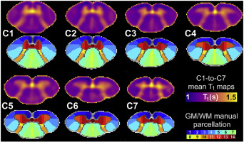
Anterior Fissure Central Canal Posterior Septum And More New Insights Into The Cervical Spinal Cord Gray And White Matter Regional Organization Using T1 Mapping At 7t Neuroimage X Mol from xpic.x-mol.com At the conus medullaris, where the spinal cord tapers, it is located more posteriorly. The space almost acts as a the human central canal of the spinal cord: 5 spinal cord this chapter briefly describes the general gross and microscopic anatomy of the spinal cord. The spinal cord forms a nearly cylindrical column that is situated within the spinal canal of the vertebral column. Curiously, with the spinal cord alone, many autonomic functions and even voluntary movements can occur. Nuclei 2 introduction to anatomy author: One at the cervical level (upper limbs), and. It is situated inside the vertebral canal of the vertebral column.
Clinical signs of spinal cord infarction include muscle weakness and paralysis with loss of reflexes.
Together with the cerebral ventricles, and the subarachnoid space of the central channel forms a single common cavity, since all. Haverkamp peter winningham winnie c. The central canal lies below and is connected to the ventricular system of the brain, from which it receives cerebrospinal fluid, and shares the same ependymal lining. The core consists mainly of two posterior (dorsal) horns, which extend toward the posterolateral surfaces of the cord, and two thicker anterior (ventral) horns, which extend toward the. Spinal cord cross section central canal. Surrounding the spinal cord and projecting downward is a slim connecting filament where the spinal cord ends (filum terminale). Gray matter (derived from embryonic mantle layer) is. Many present with serious acute symptoms such as paresthesia, paralysis, and loss of sensation or bladder and bowel function. The spinal cord forms a nearly cylindrical column that is situated within the spinal canal of the vertebral column. The central canal spans the length of the spinal cord from the caudal angle of the fourth ventricle to the conus medullaris. Spinal cord infarction (also known as a spinal stroke) refers to the death of nervous tissue, which results from an interruption of the arterial supply. The most common causes of infarction are vertebral. Central nervous system spinal cord.
It communicates with the iv ventricle and ends in a dilated region (terminal ventricle) central. Many present with serious acute symptoms such as paresthesia, paralysis, and loss of sensation or bladder and bowel function.


0 Comments